√99以上 gram stain e coli shape 269859-How to gram stain e coli
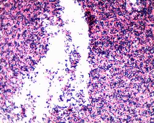
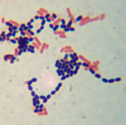
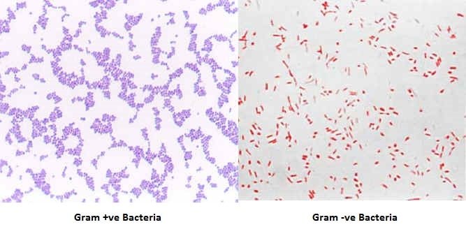
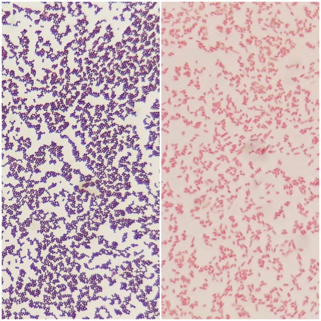
Gram stain results determine if the organism is gramnegative, but findings do not distinguish among the other aerobic gramnegative bacilli that cause similar infectious diseases E coli is aThe Gramstain is a differential stain that requires the use of at least three chemical reagents applied sequentially to a heatfixed smear The first reagent (Crystal Violet) is the primary stainIt gives color to all cells This is followed by a second reagent (Gram's Iodine), which acts as a mordant and helps the color to stick to the cell wallIn order to establish a color contrast, thisGram positive bacteria Stain dark purple due to retaining the primary dye called Crystal Violet in the cell wall Example Staphylococcus aureus Fig Gram positive bacteria 2 Gram negative bacteria Stain red or pink due to retaining the counter staining dye called Safranin Example Escherichia coli Fig Gram negative bacteria Bacterial Morphology

Laboratory Perspective Of Gram Staining And Its Significance In Investigations Of Infectious Diseases Thairu Y Nasir Ia Usman Y Sub Saharan Afr J Med
How to gram stain e coli
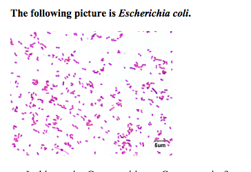
How to gram stain e coli-Bio 112 Abstract Escherichia coli, Bacillus subtilis and Staphylococcus epidermidis were analyzed for this lab activity to determine their Gram Stain After the multilayered Gram Stain procedure each bacteria were classified as Grampositive or Gramnegative depending on their cell walls staining color The results showed that E coli stained pink and classified as Gramnegative Both B subtilis and S epidermidis stained purple and were classified as Grampositive It was determined that EcYour unknown sample For example, when you perform a Gram stain, you will always include samples of Staphylococcus epidermidis (S epi), which is known to be Gram positive, and Escherichia coli, which is known to be Gramnegative If the Gram stain procedure works as it should, S epi will be purple and E coli will be pink


Laboratory Perspective Of Gram Staining And Its Significance In Investigations Of Infectious Diseases Thairu Y Nasir Ia Usman Y Sub Saharan Afr J Med
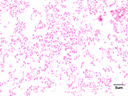
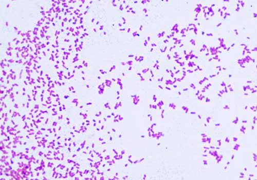
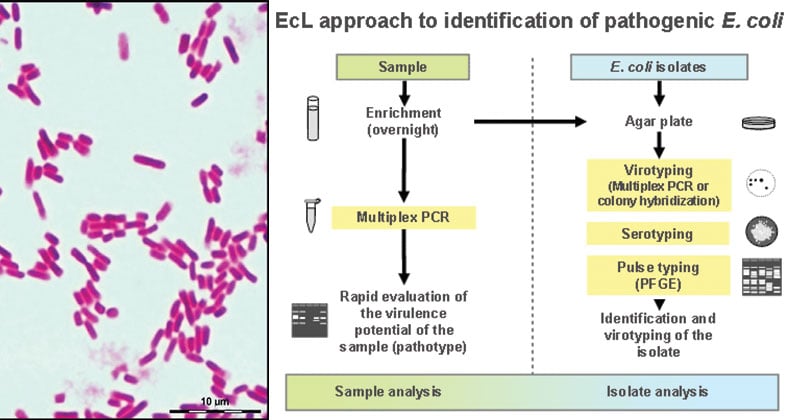
Escherichia coli Gram stained smear under microscopeRod shapedpink in colorthat's why Gram negative Bacilli#GramStain#GNB#GNRIn this work, we introduce a quantitative physical model of the bacterial cell wall that predicts the mechanical response of cell shape to peptidoglycan damage and perturbation in the rodshaped Gramnegative bacterium Escherichia coliGram Stain The Gram stain is the most important staining procedure in microbiology It is used to differentiate between gram positive organisms and gram negative organisms Hence, it is a differential stain Gram staining involves a fourpart process, which includes crystal violet, the primary stain iodine, the mordant
The Gram stain is the most widely used staining procedure in bacteriology It is called a differential stain since it differentiates between Grampositive and Gramnegative bacteria Bacteria that stain purple with the Gram staining procedure are termed Grampositive;We have cultures of E coli and Bacillus for you to gram stain This will give you gram and gram – controls to check your procedure against You can use 2 slides, 1 for each bacterium, or you can divide one slide in half and smear each bacterium on the divided slide There are also prepared, gram stained slides of bacteria of different shapes and sizes of bacteria to look atEscherichia coli Massive pyuria and numerous Gramnegative rods in a (Gram stain) Haemophilus parahaemolyticus Curved, commashaped rods in a young broth culture (Gram stain) Wautersia paucula Rod with several peritrichous flagella (Flagella stain) Yersinia enterocolitica
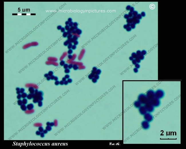
Gram positive cocci refer to Gram positive bacteria that are spherically shaped Two genera of Gram positive cocci noted for their role as human pathogens are Staphylococcus and Streptococcus Staphylococcus are spherical in shape and their cells appear in clusters after they divideSIMPLE one color only shows shape and arrangement GRAM two colors, give more information about whether they are gram or gram What color would E coli be if you used the simple stain procedure in this lab to dye it?Gramstain Grampositive cocci and Gramnegative bacilli Microscopic appearance Cocci in grapelike clusters (Saureus) and bacilli(Ecoli) Clinical significance of Saureus Frequently found as part of the normal skin flora on the skin and nasal passages;


E Coli Bacteria Britannica


2 3b The Gram Negative Cell Wall Biology Libretexts
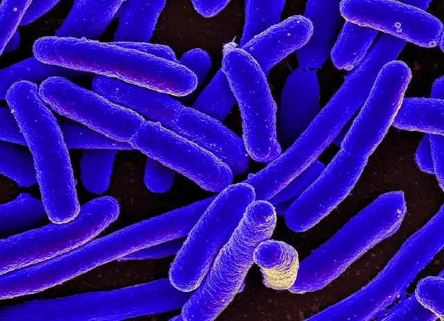
E coli serotype O157H7 is a mesophilic, Gramnegative rodshaped (Bacilli) bacterium, which possesses adhesive fimbriae and a cell wall that consists of an outer membrane containing lipopolysaccharides, a periplasmic space with a peptidoglycan layer, and an inner, cytoplasmic membraneShape – Escherichia coli is a straight, rod shape (bacillus) bacterium Size – The size of Escherichia coli is about 1–3 µm × 04–07 µm (micrometer) Arrangement Of Cells – Escherichia coli is arranged singly or in pairs Motility – Escherichia coli is a motile bacterium Some strains of E coli are nonmotileAislinn Towne February 11, 21 Experiment 4 Gram Stain Background The gram stain is one of the most used staining techniques This stain is specifically used for cellular characterization Gram positive bacteria cells can be noted by a purple color whereas Gram negative cells are a red/pink color Cells are stained through crystal violet, a mordant, then a decolorizer, and finally Safranin


Gram Stain Lab Tests Online Au


Types Of Bacteria Information From Antibiotic Research Uk
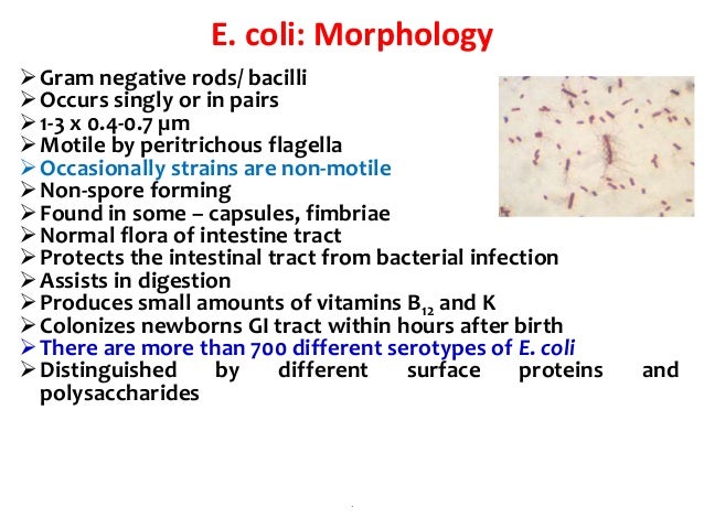
2 Morphology and Staining of Escherichia Coli E coli is Gramnegative straight rod, 13 µ x 0407 µ, arranged singly or in pairs (Fig 281) It is motile by peritrichous flagellae, though some strains are nonmotile Spores are not formed Capsules and fimbriae are found in some strains 3 Cultural Characteristics of Escherichia ColiGram staining is a quick procedure used to look for the presence of bacteria in tissue samples and to characterise bacteria as Grampositive or Gramnegative, based on the chemical and physical properties of their cell walls The Gram stain should almost always be done as the first step in diagnosis of a bacteria infection The Gram stain is named after the Danish scientist Hans ChristianAislinn Towne February 11, 21 Experiment 4 Gram Stain Background The gram stain is one of the most used staining techniques This stain is specifically used for cellular characterization Gram positive bacteria cells can be noted by a purple color whereas Gram negative cells are a red/pink color Cells are stained through crystal violet, a mordant, then a decolorizer, and finally Safranin


Pin On Microbiology


Biol 230 Lecture Guide Gram Stain Of A Mixture Of Gram Positive And Gram Negative Bacteria
2 Morphology and Staining of Escherichia Coli E coli is Gramnegative straight rod, 13 µ x 0407 µ, arranged singly or in pairs (Fig 281) It is motile by peritrichous flagellae, though some strains are nonmotile Spores are not formed Capsules and fimbriae are found in some strains 3 Cultural Characteristics of Escherichia ColiThe necks of Pasteur's flasks were bent into an Sshape and then melted to seal them After Gram staining, E coli are pink in color This result is interpreted as _____ D a, c, b C All of the following are common to both the Gram stain and the acidfast stain EXCEPT A a decolorizing agent B a decolorizing agent and aDIFFERENTIAL STAIN An example is Gram staining (or Gram's method) It is routinely used as an initial procedure in the identification of an unknown bacterial species Let's suppose we have a smear containing mixture of Staphylococcus aureus and Escherichia coli as in previous case We will use the same stains as before and besides we will need


Gram Stain Microbiology Images Photographs


Laboratory Perspective Of Gram Staining And Its Significance In Investigations Of Infectious Diseases Thairu Y Nasir Ia Usman Y Sub Saharan Afr J Med
The shape of E coli is strikingly simple compared to those of higher eukaryotes Outwardly, it resembles a lower eukaryote like S pombe However, the latter not only is larger (7 to 13 μm long and 2 to 3 μm in diameter versus 15 to 55 μm long and 05 to 10 μm in diameter) but also harbors membranebound organelles and a cytoskeletonBiology Of E Coli E coli (Escherichia coli) are a small, Gramnegative species of bacteria Most strains of E coli are rodshaped and measure about μm long and 0210 μm in diameter They typically have a cell volume of 0607 μm, most of which is filled by the cytoplasmRESULTS After completing the experiment, it was concluded that M luteus is gram positive and E coli and S marcescens are gram negative As indicated in the student notes, the first slide that was prepared using aseptic technique and the Gram Staining method was a slide containing cultures of M luteus and E coli When examining this slide under the microscope at 1000X magnification, I observed some clusters and mostly doubles of coccishaped purple colonies


Asmscience Examination Of Gram Stains Of Urine


Identifying Bacteria Through Look Growth Stain And Strain
SUMMARY The shape of Escherichia coli is strikingly simple compared to those of higher eukaryotes In fact, the end result of E coli morphogenesis is a cylindrical tube with hemispherical caps It is argued that physical principles affect biological forms In this view, genes code for products that contribute to the production of suitable structures for physical factors to act uponGram Staining Now that the slide has been heat fixed, you may now begin to stain the organism This is started by applying a few drops of crystal violet for 30 seconds, then rinsing with water for 5 seconds Next, cover the slide with Gram's Iodine and let sit for a full minute before rinsing with water for another 5 secondsFor the rodshaped Gramnegative bacterium Escherichia coli, changes in cell shape have critical consequences for motility, immune system evasion, proliferation and adhesion For most bacteria, the peptidoglycan cell wall is both necessary and sufficient to determine cell shape However, how the synthesis machinery assembles a peptidoglycan network with a robustly maintained micronscale shape has remained elusive


Solved Lab Module Staining Study Help Instructions Rea Chegg Com


Growth Requirements Of E Coli And Auxotrophs Microbiology Class Video Study Com
Most of the bacteria range from 022 µm in diameter The length can range from 110 µm for filamentous or rodshaped bacteria The most wellknown bacteria E coli, their average size is ~15 µm in diameter and 26 µm in length In this figure The size comparison between our hair (~ 60 µm) and E coli (~1 µm) Notice how small the bacteria areEnterotoxigenic Escherichia coli Overview Enterotoxigenic Escherichia coli (ETEC) is a Gramnegative, rodshaped bacterium that causes traveler's diarrhea (Figure 1)ETEC bacteria are motile facultative anaerobes (they can makes ATP energy by aerobic respiration if oxygen is present, but is also capable of switching to fermentation) that can easily be cultured under aerobic conditions at 37°CSo the Gram stain, when you first apply it to a bacteria, it just stains the whole thing purple, and then you'll wash it off And because the Gram negative bacteria has this very thin peptidoglycan layer in their cell wall, it washes right off, and later they'll restain it with something called Safranin, which isn't important, but they come in


E Coli Under The Microscope Types Techniques Gram Stain Hanging Drop Method


Sem Images Of Gram Negative Bacteria Including E Coli And P Download Scientific Diagram
Most of the bacteria range from 022 µm in diameter The length can range from 110 µm for filamentous or rodshaped bacteria The most wellknown bacteria E coli, their average size is ~15 µm in diameter and 26 µm in length In this figure The size comparison between our hair (~ 60 µm) and E coli (~1 µm) Notice how small theEscherichia coli (commonly abbreviated E coli) is a Gramnegative, rodshaped bacterium that is commonly found in the lower intestine of warmblooded organisms (endotherms) Most E coli strains are harmless, but some serotypes can cause serious food poisoning in humansIt is estimated that % of the human population are longterm carriers of S aureus


Welcome To Microbugz Gram Stain


Gram Staining Principle Procedure Interpretation Examples And Animation
If you only had one dye, all bacteria on the slide will look blue This is different from the Gram result where E coliThose that stain pink are said to be Gramnegative The terms positive and negative have nothing to do with electrical charge, but simply designate two distinct morphological groups of bacteriaThe Gram stain is the most widely used staining procedure in bacteriology It is called a differential stain since it differentiates between Grampositive and Gramnegative bacteria Bacteria that stain purple with the Gram staining procedure are termed Grampositive;


1


Bacterial Morphotypes In Sputum Gram Stain 100 Oil Immersion Field Download Scientific Diagram
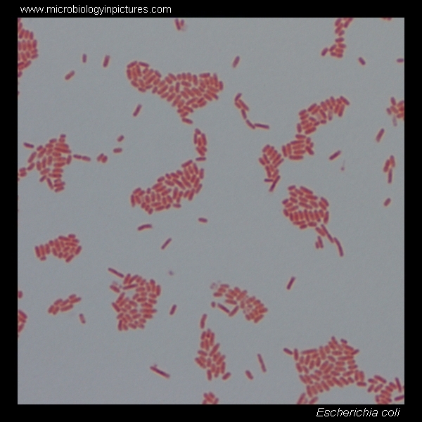
Gram stain results determine if the organism is gramnegative, but findings do not distinguish among the other aerobic gramnegative bacilli that cause similar infectious diseases E coli is aThose that stain pink are said to be Gramnegative The terms positive and negative have nothing to do with electrical charge, but simply designate two distinct morphological groups of bacteriaWhen viewed under the microscope, Gramnegative E Coli will appear pink in color The absence of this (of purple color) is indicative of Grampositive bacteria and the absence of Gramnegative E Coli


E Coli Gram Stain Introduction Principle Procedure And Result Interpret


Gram Negative Bacteria Images Photos Of Escherichia Coli Salmonella Enterobacter
Gram positive cocci refer to Gram positive bacteria that are spherically shaped Two genera of Gram positive cocci noted for their role as human pathogens are Staphylococcus and Streptococcus Staphylococcus are spherical in shape and their cells appear in clusters after they divideEscherichia coli Gram stained smear under microscopeRod shapedpink in colorthat's why Gram negative Bacilli#GramStain#GNB#GNRGram stain or Gram staining, also called Gram's method, is a method of staining used to distinguish and classify bacterial species into two large groups grampositive bacteria and gramnegative bacteriaThe name comes from the Danish bacteriologist Hans Christian Gram, who developed the technique Gram staining differentiates bacteria by the chemical and physical properties of their cell walls


Gram Stain Staphylococcus Aureus And Escherichia Coli Gram Staining Technique Micrograph Of S Aureus And E Coli


Gram Positive Cocci An Overview Sciencedirect Topics
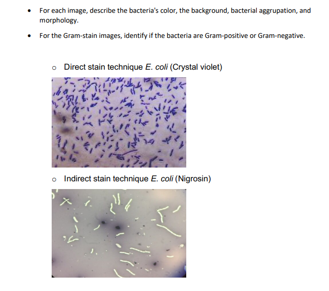
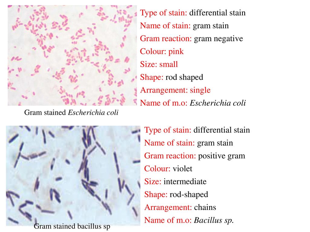
E coli is a gramnegative, rodshaped, facultative anaerobic bacterium that belongs to family Enterobacteriaceae It is a faecal coliform bacterium commonly found in the lower intestine of warmblooded organisms Many E coli strains are harmless, and they are a part of the normal microbiota of the gut that keeps the gut healthy But, some serotypes cause serious food poisoning, severe abdominal cramps, bloody diarrhoea, kidney failure and vomiting2 Morphology and Staining of Escherichia Coli E coli is Gramnegative straight rod, 13 µ x 0407 µ, arranged singly or in pairs (Fig 281) It is motile by peritrichous flagellae, though some strains are nonmotile Spores are not formed Capsules and fimbriae are found in some strains 3 Cultural Characteristics of Escherichia ColiBacteria Escherichia coli Shape Rod shaped Disease Ecoli infection, diarrhea, abd pain Notes Gram stain is used to determine size, shape, and arrangment As well as gram positive or negative Color Red color for negative Len 100x;


Phenotypic Plasticity Of Escherichia Coli Upon Exposure To Physical Stress Induced By Zno Nanorods Scientific Reports


Gram Stain Of E Coli Bacterium A Gram Stain Of Shows Gramnegative Download Scientific Diagram
In this work, we introduce a quantitative physical model of the bacterial cell wall that predicts the mechanical response of cell shape to peptidoglycan damage and perturbation in the rodshaped Gramnegative bacterium Escherichia coliE coli are classified as facultative anaerobes, which means that they grow best when oxygen is present but are able to switch to nonoxygendependent chemical processes in the absence of oxygen E coli bacteria are gramnegative, so they stain pink in a gram test E coli bacteria were first identified by bacteriologist Dr Escherich in 15Muscle biopsy examined under the microscope (haematoxylineosin stain, zoom 100×) the large white areas between the muscle fibers are due to gas formation Gram stain of a muscle biopsy showing Grampositive , rodshaped , anaerobic , sporeforming bacteria in the infected muscle tissue The result is highly compatible with an infection with C perfringens


Escherichia Coli E Coli An Overview Microbe Notes


Solved For Each Image Describe The Bacteria S Color The Chegg Com
Escherichia coli ( / ˌɛʃəˈrɪkiə ˈkoʊlaɪ / ), also known as E coli ( / ˌiː ˈkoʊlaɪ / ), is a Gramnegative, facultative anaerobic, rodshaped, coliform bacterium of the genus Escherichia that is commonly found in the lower intestine of warmblooded organisms (endotherms)The two classes of bacteria are differentiated through gram staining, because of their cell wall composition ie Grampositive bacteria have a large layer of peptidoglycan and a thin layer of the lipid layer, and unlike the Gramnegative bacteria which have a thick layer of lipids and they lack the peptidoglycan layer or some have a very thin layer of the peptidoglycan layerEscherichia coli (E coli) bacteria are bacillus shaped bacteria Most strains of E coli that reside within us are harmless and even provide beneficial functions, such as food digestion, nutrient absorption, and the production of vitamin K Other strains, however, are pathogenic and can cause intestinal disease, urinary tract infections, and


Gram Positive Bacteria Microbiology


Escherichia Coli Bacteria Appearance Dangerous Infections And Diseases E Coli O157 H7 Treatment Organs And Body Parts Affected By E Coli O157 H7 Bacteria Cell Morphology And Gram Stain Gram Negative Bacteria
What is E Coli?Escherichia coli (/ ˌ ɛ ʃ ə ˈ r ɪ k i ə ˈ k oʊ l aɪ /), also known as E coli (/ ˌ iː ˈ k oʊ l aɪ /), is a Gramnegative, facultative anaerobic, rodshaped, coliform bacterium of the genus Escherichia that is commonly found in the lower intestine of warmblooded organisms (endotherms) Most E coli strains are harmless, but some serotypes (EPEC, ETEC etc) can cause serious foodYour unknown sample For example, when you perform a Gram stain, you will always include samples of Staphylococcus epidermidis (S epi), which is known to be Gram positive, and Escherichia coli, which is known to be Gramnegative If the Gram stain procedure works as it should, S epi will be purple and E coli will be pink
/gram-positive-staphylococcus-aureus-bacteria-541802136-57979cca5f9b58461f26eccc.jpg)


Gram Stain Procedure In Microbiology


Capsule Staining Principle Reagents Procedure And Result


Gram Negative Bacteria Shape Page 4 Line 17qq Com


Gram Stain Images Microbiology Stain Study Tools


Optical Microscope Images Of E Coli Cells Following Gram Staining A Download Scientific Diagram


E Coli Gram Stain And Cell Morphology E Coli Micrograph Appearance Under The Microscope E Coli Cell Morphology E Coli Microscopic Picture


E Coli Why So Famous


The Lab Freaks Testing On Escherichia Coli
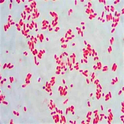


Morphology Culture Characteristics Of Escherichia Coli E Coli


Academic Oup Com Labmed Article Pdf 32 7 368 Labmed32 0368 Pdf
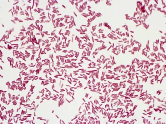


Biochemical Test Of Escherichia Coli E Coli Microbe Notes


Cell Shape And Cell Wall Organization In Gram Negative Bacteria Pnas


Figure 3 From Pathologicalstudy On Colibacillosis In Chickens And Detection Of Escherichia Coli By Pcr Semantic Scholar


Gram Stain Of E Coli Bacterium A Gram Stain Of Shows Gramnegative Download Scientific Diagram
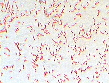


Photo Gallery Of Pathogenic Bacterial
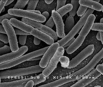


Escherichia Coli Microbewiki


Q Tbn And9gctnncfjtcedl5fo0rq5mdljtvlng Qopeaabn2fkjwz27muvuqg Usqp Cau


Q Tbn And9gcqkye60ou Johpr02n Mbv1fferrjpdh Lnct7ymdf5qhyia1ld Usqp Cau


1b Demo


Www Wiv Isp Be Qml Activities External Quality Rapports Atlas Bacteriology Gram Negative Aerobic And Facultative Rods Pdf


Solved 1 Identify The Morphology Morphological Arrangem Chegg Com
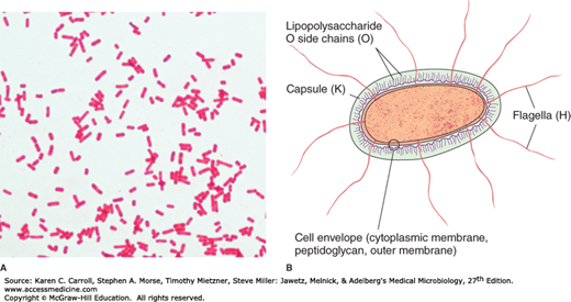


Enteric Gram Negative Rods Enterobacteriaceae Basicmedical Key


Gram Stain Wikipedia


Gram Negative Rods Ppt Download


Morphology Of Bacterial Cells


Salmonella And Salmonellosis


Q Tbn And9gctgrmjg8acjrbxbniknzy1qewngtoobg8cixbwsv Ok9ny02ti0 Usqp Cau


Staining Microscopic Specimens Microbiology


Genus Escherichia Coli


Academic Oup Com Labmed Article Pdf 32 7 368 Labmed32 0368 Pdf


Laboratory Science Review Microbiology Medical Laboratory Science Medical Laboratory


Solved Gram Stain Results Below Are Images Of Bacteria I Chegg Com


Gram Staining Principle Procedure And Results Msc Sarah Ahmed Ppt Download


Pin On Microcosmos


Pathogens Bacteria Viruses Gram Stain Teachmephysiology


Gram Stain


Bacteria Diversity Of Structure Of Bacteria Britannica


Laboratory Perspective Of Gram Staining And Its Significance In Investigations Of Infectious Diseases Thairu Y Nasir Ia Usman Y Sub Saharan Afr J Med


Bacilli Wikipedia


What Does An E Coli Bacteria Look Like Under A Microscope Quora
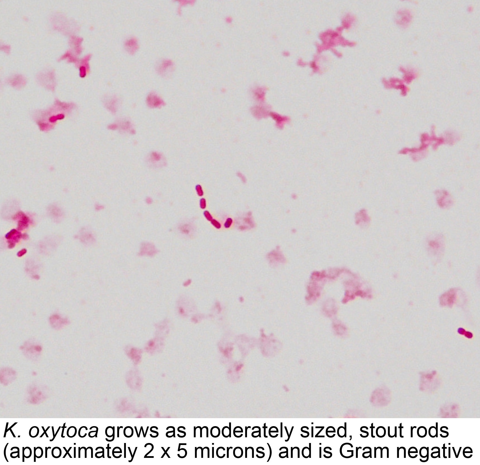


Pathology Outlines Klebsiella Oxytoca


Http Article Sciencepublishinggroup Com Pdf 10 J Fem 13 Pdf
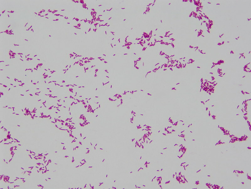


Pulsenotes Gram Negative Infections Notes


Bacterial Staining


Escherichia Coli Colony Morphology And Microscopic Appearance Basic Characteristic And Tests For Identification Of E Coli Bacteria Images Of Escherichia Coli Antibiotic Treatment Of E Coli Infections


Bacteria 101 Cell Walls Gram Staining Common Pathogens Tusom Pharmwiki
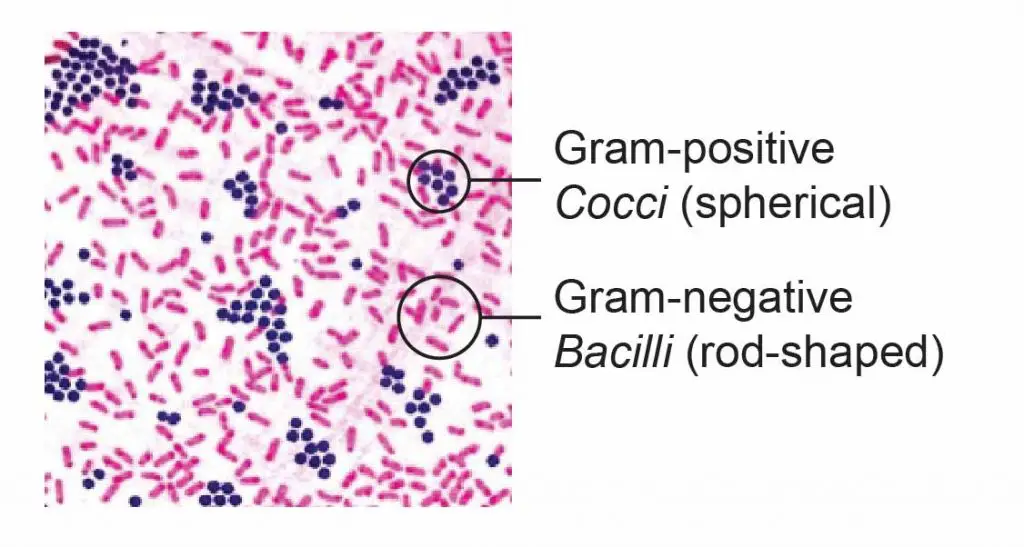


Observing Bacteria Under The Microscope Gram Stain Steps Rs Science


Optical Microscope Images Of E Coli Cells Following Gram Staining A Download Scientific Diagram


Colony Characteristics Of E Coli Sciencing


Escherichia Coli Wikipedia
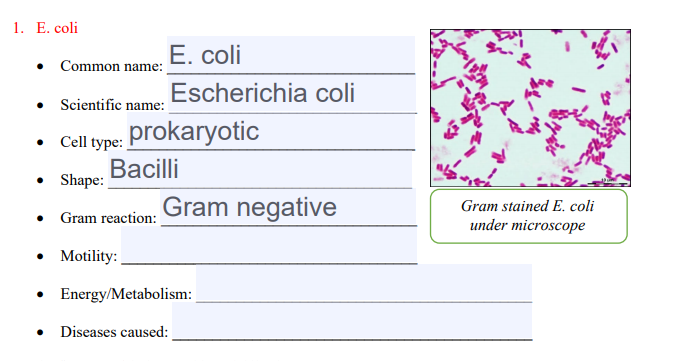


Solved 1 E Coli E Coli Common Name Escherichia Coli S Chegg Com


Vibrio Cholerae In Gram Stain Introduction Procedure And Result Interpret


Solved 1 Identify The Morphology Morphological Arrangem Chegg Com


Chapter 2 Cells


Effect Of The Compound No On Cell Morphology Of E Coli Cells Download Scientific Diagram


What Is The Cell Morphology Of Escherichia Coli Quora


Escherichia Coli Wikipedia


Gram Negative Bacteria Wikipedia


Bacterial Characteristics Gram Staining Video Khan Academy
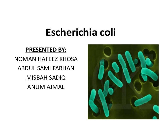


E Coli
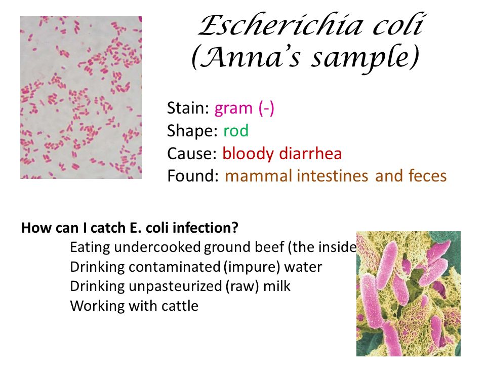


What Should You Have Seen Under The Microscope Activity Ppt Download


Module 7 8 10 Gram Stain Acid Fast Endospore Growth Characteristics Flashcards Quizlet


E Coli In Gram Stain Introduction Pathogenic Strains And Lab Diagnosis


Www Mccc Edu Hilkerd Documents Bio1lab3 Exp 4 000 Pdf
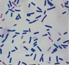


Gram Staining Rules


E Coli Gram Stain Mikrobiologiya


Staphylococcus Aureus And Ecoli Under Microscope Microscopy Of Gram Positive Cocci And Gram Negative Bacilli Morphology And Microscopic Appearance Of Staphylococcus Aureus And E Coli S Aureus Gram Stain And Colony Morphology On Agar Clinical
/gram_positive_vs_negative-5b7f26d2c9e77c005746fbd7.jpg)


Gram Positive Vs Gram Negative Bacteria
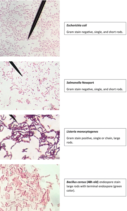


Staining Technology And Bright Field Microscope Use Springerlink


Escherichia Coli E Coli Meaning Morphology And Characteristics


Enterococcus Faecalis An Overview Sciencedirect Topics
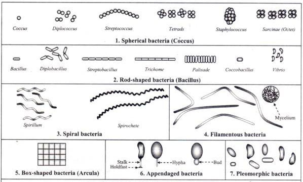


Different Size Shape And Arrangement Of Bacterial Cells
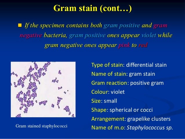


Bacterial Staining


Recombinant Production Of The Therapeutic Peptide Lunasin Microbial Cell Factories Full Text


What Does Gram Positive Mean Fermup
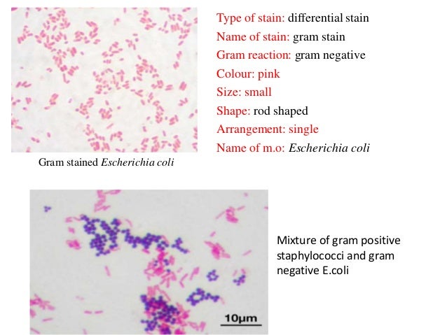


Bacterial Staining


コメント
コメントを投稿